GFP-Booster

7
Description
anti-GFP VHH/ Nanobody conjugated to a fluorescent dye for IF/ microscopy.
Conjugations
Alexa Fluor® 488
Alexa Fluor® 568
Alexa Fluor® 647
ATTO488
ATTO594
ATTO647N
Unconjugated
Specificity
CFP, eGFP, AcGFP, mClover (Clover A206K), eYFP, Venus and more GFP derivatives, see:
Fluorescent protein specificity table Nano-Booster
Applications
Immunofluorescence (IF): Immunohistochemistry (IHC), Immunocytochemistry (ICC)
Wide-field fluorescence and confocal microscopy, super-resolution microscopy (SRM), light-sheet microscopy
Cleared tissue, organ, and animal imaging
Product | Size | Code | Price | Buy |
---|---|---|---|---|
Product GFP-Booster Alexa Fluor® 488 | Size 10 µL | Code gb2AF488-10 | Price $ 170 |
Buy
![]() |
Product GFP-Booster Alexa Fluor® 488 | Size 50 µL | Code gb2AF488-50 | Price $ 395 |
Buy
![]() |
Product GFP-Booster Alexa Fluor® 568 | Size 10 µL | Code gb2AF568-10 | Price $ 170 |
Buy
![]() |
Product GFP-Booster Alexa Fluor® 568 | Size 50 µL | Code gb2AF568-50 | Price $ 395 |
Buy
![]() |
Product GFP-Booster Alexa Fluor® 647 | Size 10 µL | Code gb2AF647-10 | Price $ 170 |
Buy
![]() |
Product GFP-Booster Alexa Fluor® 647 | Size 50 µL | Code gb2AF647-50 | Price $ 395 |
Buy
![]() |
Product GFP-Booster Atto488 | Size 10 µL | Code gba488-10 | Price $ 100 |
Buy
![]() |
Product GFP-Booster Atto488 | Size 100 µL | Code gba488-100 | Price $ 365 |
Buy
![]() |
Product GFP-Booster Atto594 | Size 10 µL | Code gba594-10 | Price $ 100 |
Buy
![]() |
Product GFP-Booster Atto594 | Size 100 µL | Code gba594-100 | Price $ 365 |
Buy
![]() |
Product GFP-Booster Atto647N (please inquire) | Size 10 µL | Code gba647n-10 | Price $ 110 |
Buy
![]() |
Product GFP-Booster Atto647N (please inquire) | Size 100 µL | Code gba647n-100 | Price $ 395 |
Buy
![]() |
Product GFP VHH, recombinant binding protein | Size 250 µL | Code gt-250 | Price $ 275 |
Buy
![]() |
Product GFP-Binding protein, custom labeling | Size | Code custom | Price Please inquire |
Coupled Nanobody/ VHH
Recombinant, monoclonal anti-Green Fluorescent Protein (GFP) single domain antibody (sdAb) fragment
Specificity
AcGFP, Citrine, CFP, eGFP, eYFP, GFP S65T, pHluorin, sfGFP, mClover (Clover A206K), TagGFP, tagGFP2, Venus, wtGFP, YFP
Host/ isotype
Alpaca/recombinant VHH domain, monoclonal
Available conjugates
Alexa Fluor® 488, Alexa Fluor® 568, Alexa Fluor® 647, ATTO488, ATTO594, ATTO647N, unconjugated
Recommended dilution for GFP-Booster Alexa Fluor® 488, Alexa Fluor® 568, Alexa Fluor® 647
IF/ICC/IHC: 1:500- 1:1,000 Optimal working concentration is application-dependent and should be determined by testing a range of dilutions from 1:100 to 1:4,000
Recommended dilution for GFP-Booster ATTO488, ATTO594, ATTO647N
IF/ICC/IHC: 1:200 Optimal working concentration is application-dependent and should be determined by testing a range of dilutions from 1:50 to 1:1,600
Microscopy techniques
Wide-field epifluorescence microscopy; confocal microscopy; super-resolution microscopy e.g. 3D-SIM, PALM, STED, STORM; light-sheet microscopy
Form
Purified recombinant protein in PBS supplemented with preservative 0.09 % sodium azide
Size
10 μL, 50µL, 100 μL
Because of the small size of a VHH these volumes contain about 10 times more molecules than a comparable volume of conventional IgG antibodies.
Protein concentration
0.5 – 1 mg/mL (conjugates)
RRID
GFP-Booster Alexa Fluor 488: AB_2827573
GFP-Booster Alexa Fluor 568: AB_2827574
GFP-Booster Alexa Fluor 647: AB_2827575
GFP-Booster Atto488: AB_2631386
GFP-Booster Atto594: AB_2631387
GFP-Booster Atto647N: AB_2629215
GFP-Booster Abberior AS 635P: AB_2631388
Storage instructions
Shipped at ambient temperature. Protect from light. For storage instructions see Manual/ Data Sheet.
Validation
Validated in cell culture & cell lines, tissue sections, yeast, fly, zebrafish, mouse, in germline of Caenorhabditis elegans, in Drosophila melanogaster embryos and cleared whole mouse. Fixed cultured cells: formaldehyde, methanol or glutaraldehyde fixation
Tissue sections: cryosections, FFPE paraffin sections
Whole specimen: cleared mouse
Affinity
Dissociation constant KD of 1 pM, detected using switchSENSE® technology from Dynamic Biosensors
Protocol
- Protocol GFP-Booster ATTO488 (PDF)
- Protocol GFP-Booster ATTO594 (PDF)
- Protocol GFP-Booster ATTO647N (PDF)
- Protocol GFP-Booster Alexa Fluor 488 (PDF)
- Protocol GFP-Booster Alexa Fluor 568 (PDF)
- Protocol GFP-Booster Alexa Fluor 647 (PDF)
- Optimized protocol for yeast (Kaplan & Ewers)
- Dual-color STORM protocol (Bleck et al.)
- Dual-color STORM protocol (Platonova et al.)
SDS
- SDS GFP-Booster ATTO488 (PDF)
- SDS GFP-Booster ATTO594 (PDF)
- SDS GFP-Booster ATTO647N (PDF)
- SDS GFP-Booster Alexa Fluor 488 (PDF)
- SDS GFP-Booster Alexa Fluor 568 (PDF)
- SDS GFP-Booster Alexa Fluor 647 (PDF)
Brochure
Which Nano-Booster and Nano-Label conjugates are recommended for super-resolution microscopy?
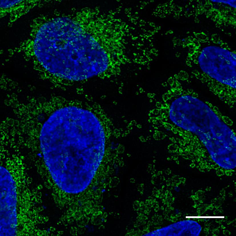
Nano-Boosters and Nano-Labels are highly suitable for Super-Resolution Microscopy. Due to their small size (2-3 nm), they minimize the linkage error and provide a more precise and dense staining than conventional antibodies (15 nm linear dimension). The selection of a Nano-Booster and Nano-Label conjugate depends on your microscope setup and lasers. We recommend for:
- STED: ATTO647N, Abberior STAR 635P
- STORM: Alexa Fluor 647, ATTO488
- SIM: ATTO488/594
Are Nano-Booster applicable for live-cell imaging?
Yes, if the fusion-tag is on the cell surface.
Nano-Boosters and Nano-Labels are small proteins and therefore don’t penetrate through non-permeabilized cell membranes. Hence, if your fusion-protein is intracellular, you may want to apply protein transduction methods (e.g. electroporation) or reagents, however from our experience, the most efficient way is to microinject the Nano-Boosters and Nano-Labels.
Can I do a simultaneous co-staining with two or more Nano-Boosters and Nano-Labels?
Yes, you can combine the Nano-Boosters and Nano-Labels. For example, if you typically use the Nano-Boosters in a 1:200 dilution, you should add 1 µL each of gba488 and rba594 to 200 µL of blocking solution for a co-staining.
How many dye molecules are coupled to Nano-Boosters and Nano-Labels?
Each Nano-Booster and Nano-Label molecule carries on average 1-2 fluorophores. Nano-Boosters conjugated to Alexa Fluor® dyes are labeled in a site-directed way and carry in total 2 fluorophores per VHH. Nano-Boosters labeled with Atto647N carry a maximum of 1 fluorophore per VHH at the C-terminus.
Can I do two-color super-resolution microscopy combining GFP- and RFP-Boosters?
Yes, dual-color STORM with Nano-Boosters is described in Bleck et al., PNAS 2014 and Platonova et al., ACS Chem Biol 2015 .
Do Nano-Boosters work on (methanol-) fixed samples?
Yes. Nano-Booster stainings perform equally well after fixation with most common reagents: paraformaldehyde, glutaraldehyde, methanol (Kaplan & Ewers, 2015; Ries et al., 2012).
What is the protocol for live-cell Nano-Booster and Nano-Label staining of the extracellular fusion protein?
Incubate the cells with 1:25 Nano-Booster or Nano-Label in growth media for 15 min at +4°C, wash and image. This protocol will highlight just the plasma membrane pool of your fusion protein.
Do Nano-Boosters and Nano-Labels penetrate though the cell membranes of live cells?
No. Nano-Boosters and Nano-Labels are small proteins and therefore don’t penetrate through non-permeabilized cell membranes. If you need to deliver Nano-Booster and Nano-Labels into live cells, you may want to apply protein transduction methods (e.g. electroporation) or reagents, however from our experience, the most efficient way is to microinject the Nano-Boosters and Nano-Labels.
Is it possible to conjugate Nano-Boosters and Nano-Labels to other fluorophores?
Yes. You can label the ChromoTek GFP-Binding Protein (GFP VHH, product code: gt-250), RFP-Binding Protein (RFP VHH, product code: rt-250) and Spot-Binding Protein (Spot VHH, product code: etb-250) with NHS-activated fluorescent dyes following the instructions of the dye manufacturer.
Note: Spot VHH contains a sortase-tag at its C terminus (sortase recognition motif LPETG) which can be used for conjugation.
Can I do IF in yeast with Nano-Boosters?
Yes, immunostaining of yeast with Nano-Boosters is in fact simpler than with traditional (IgG) antibodies, because Nano-Boosters can penetrate the yeast cell wall due to their small size. For an optimized protocol for yeast staining with Nano-Boosters (here a GFP nanobody) see Kaplan & Ewers, Nat Protoc. 2015.
GFP-Booster Alexa Fluor or GFP-Booster ATTO
We currently offer our GFP-Booster conjugated to different Alexa Fluor dyes and to several ATTO fluorophores.
The GFP-Boosters conjugated to Alexa Fluor have multiple advantages, when compared to GFP-Boosters labelled with ATTO dyes:
Improved GFP-Booster Alexa Fluor | GFP-Booster ATTO |
Brighter fluorescence signal | About one third as bright |
Site directed labelled | Randomly labelled |
Degree of labeling DOL = 2 dyes per VHH | Labelled with average 1 fluorophore per VHH |
Dilution range: 1:500 – 1:1,000 | Dilution range: 1:200 |
Product codes: gb2AF488, gb2AF568, gb2AF647 | Product codes: gba488, gba594, gba647n |
Note that we intend to replace GFP-Boosters ATTO by GFP-Booster Alexa Fluor.
Please contact us if you require an ongoing supply of GFP-Boosters conjugated to ATTO dyes. We are curious to learn why you need GFP-Booster ATTO and why you can’t use GFP-Boosters Alexa Fluor instead.
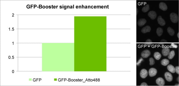
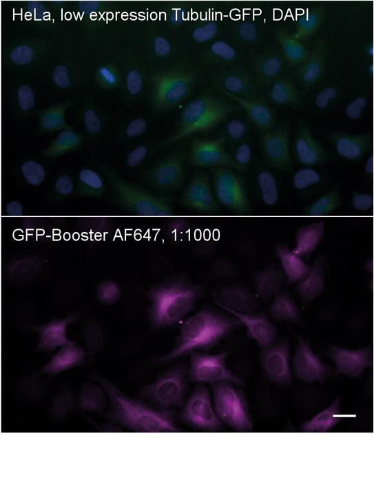
Stabilization, enhancement and reactivation of fluorescent protein signals with GFP-Booster
Fluorescent proteins (FPs) are powerful tools to study protein localization and dynamics in living cells. However, genetically encoded FPs have several disadvantages compared to chemical dyes:
- Signal intensities of fixed samples from cells expressing FP fusion at physiological expression levels are usually very low.
- Both photostability and quantum efficiency of FPs are generally not sufficient for super-resolution microscopy (e.g. 3D-SIM, STED or STORM/PALM).
- Many cell biological methods such as HCl treatment for BrdU-detection, the EdU-Click-iT™ treatment or heat denaturation for FiSH lead to disruption of the FP signal.
Here, the ChromoTek GFP-Booster will help you to get better images from your existing GFP expression constructs.
Benefits
- Considerably higher tissue penetration rates
- Superior accessibility and labelling of epitopes in crowded cellular/organelle environments
- Less than 2 nm epitope-label displacement minimizes linkage error
- Monovalent VHHs do not cluster their epitopes
- Validation: structure and function characterized
- Consistent and reliable performance due to recombinant production
Only for research applications, not for diagnostic or therapeutic use!